Unit Iv Soft Tissue Mechanics The Soft Tissues of The Body Kripas Biology Diagrams
Unit Iv Soft Tissue Mechanics The Soft Tissues of The Body Kripas Biology Diagrams Start studying Soft Tissue Anatomy. Learn vocabulary, terms, and more with flashcards, games, and other study tools. They consist of three different hard, or mineralized, tissues - dentin, cementum, and enamel - and are supported by a fourth hard tissue - bone (Fig. 1.5). The interface between the teeth and the gingivae is the only place in the body where a structure composed of hard tissues breaches a soft tissue covering.

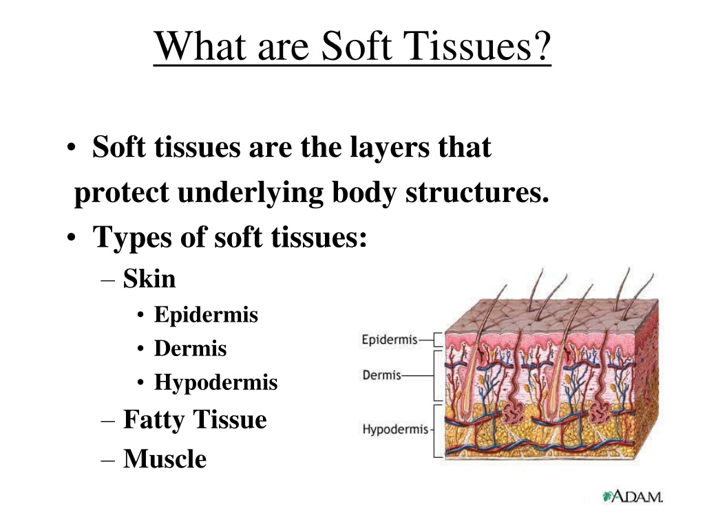
The layers of the skin make up the body's largest organ, providing a crucial barrier between the internal structures and the external environment. This complex, multi-layered tissue is essential for protection, sensation, temperature regulation, and immune defense. Understanding the structure and function of the layers of the skin is key to appreciating its role in human health and physiology.
4.1 Types of Tissues Biology Diagrams
Supportive connective tissue—bone and cartilage—provide structure and strength to the body and protect soft tissues. A few distinct cell types and densely packed fibers in a matrix characterize these tissues.

Rectus Capitis Muscle Sternocleidomastoid Muscle Palantine Tonsil Soft Palate Retromandibular Vein Maxillary Tuberosity Vertebral Artery in Transverse Foramin

Soft Tissue Anatomy Diagram Biology Diagrams
File:DIagram of the different types of soft tissue in the body CRUK 037.svg Download scientific diagram | Soft tissue anatomy from publication: Soft Tissue Modelling from 3D Scanned Data | Human body 3D scanners are becoming a mature technology that generates accurate

Download scientific diagram | Anatomy of skin and soft tissue structures and layers commonly involved with various infectious processes. from publication: Treatment of Complicated Skin and Soft
